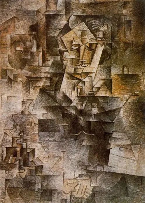
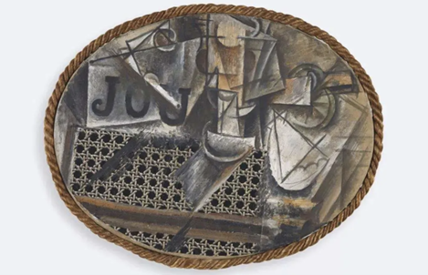
At the beginning of the 20th century, a unique art school called “Cubism” emerged in Paris, France, and one of its masters was the well-known Pablo Picasso. Artists look at all sides of an object, and then, with just a flat drawing, they are able to represent all of them at the same time. However, since different objects and faces are intertwined in the same plane, these paintings give an intuitive feeling of ” abstract ” and ” difficult to identify “.

Picasso analyzes the Cubist period (1907-1911) work “Kassler”
Today, we often need to use spectral imaging in research in remote sensing satellites, biomedicine, and more.
The multi-band images captured by remote sensing satellites contain signals on different spectral channels, and different types of vegetation and geological structures correspond to characteristic spectral distributions; the same is true for the emission spectra of fluorophores when imaging biomolecules. The spectral image we end up with is a mixture of contributions from all fluorophores (staining molecules) in the observed area, and if more than four fluorophores are used, the colors they produce will overlap and be indistinguishable; as in the period of analytical cubism, Different angles of objects are compressed into the same picture by Picasso.
After entering the period of Synthetic Cubism, Picasso began to use disintegrated abstract graphics to construct the form of real objects in turn. Corresponding to our current spectroscopy, it is like unmixing of the spectrum (unmixing, that is, the decomposition of the mixed spectrum) – to resolve the contributions of different channels/fluorophores within the same pixel.

Picasso’s Statue of Kathler during the Synthetic Cubism Period (1912-1914)
Historically, unmixing of spectra has typically relied on linear decomposition, describing the fluorescence signal as a linear mixture of contributions from all fluorophores in the observed region. This requires the reference spectra of all relevant fluorophores, ie the spectra of a single fluorophore. The emission spectrum of each subregional fluorophore needs to be measured separately, so making reference spectral measurements in a highly heterogeneous specimen like the brain is often a complex, inefficient, and time-consuming process.
Recently, a research team from the Korea Institute of Science and Technology (KAIST) published a new imaging method named after Picasso in the journal Nature Communication . Co-corresponding author Young-Gyu Yoon, a professor in the School of Electrical Engineering at KAIST, said, “To solve the problem of difficult access to reference spectra, we developed a method that does not require measurement of reference spectra .” Using artificial intelligence, they made a breakthrough in 4-color imaging , and the reliance on reference spectra of past linear unmixing techniques, the ability to use more than 15 colors to image and resolve spatially overlapping proteins or other biomolecules can elucidate even the most complex subject: the brain.

webpage Screenshot
The full name of the PICASSO method is “Process of ultra- multiplexed Imaging of biomolecules viA the unmixing of the Signals of Spectrally Overlapping fluorophores”. “Super multiplexed imaging” refers to the visualization of many individual components of a unit. Just like every movie theater in a movie theater plays a different movie, every protein in a cell has a different role, and fluorophore staining is an important way to study these different roles.
“We devised an information theory-based strategy to unmix through an iterative approach that minimizes repeated information between multiple channels in a mixed image,” said co-corresponding author Jae-Byum Chang, a professor in the Department of Materials Science and Engineering at KAIST. , “This allows us to get rid of the assumption that the spatial distributions of different proteins are mutually exclusive, and achieve accurate information unmixing.”
To demonstrate the capabilities of PICASSO, the researchers performed a single round of staining on mouse brains and performed multiplexed imaging of 15 colors using the technique. The mouse brain, despite its small size, is still very complex, and if only other imaging techniques such as circulating immunofluorescence are used, it may require extensive re-staining and a significant investment of time, manpower and resources. However, combining the circulating immunofluorescence technique with PICASSO, the research team achieved 45-color multiplexed imaging of the mouse brain in just three staining-imaging cycles.

Results of fifteen-color multiplexed imaging of mouse brain by PICASSO (a is the emission spectrum of the 15 fluorophores used. b Fifteen-color multiplexed imaging of the dentate gyrus of the mouse hippocampus. cq are the different target proteins and their corresponding A single-channel image of b) | Reference [1]
“PICASSO is a versatile tool that can be used for multiplexed biomolecular imaging of cells, tissue sections and clinical specimens,” Chang said. “We expect PICASSO to have a wide range of applications. In terms of spatial information of biomolecules, this technology It will help to reveal the tumor microenvironment, especially the cellular heterogeneity of the heterogeneous population of immune cells, which is also closely related to cancer prognosis and the efficacy of cancer therapy. At the same time, PICASSO is also expected to enable 3D visualization of larger biomolecular populations , providing information on the co-localization and expression of different proteins or mRNA molecules.”
references
[1]Chang, J. (2022, May 5). PICASSO allows ultra-multiplexed fluorescence imaging of spatially overlapping proteins without reference spectra measurements. Nature. https://ift.tt/9O2YiCd
[2] https://ift.tt/GaQ0NK1
Compilation: Bamboo
Editor: Jin Xiaoming
Typesetting: Yin Ningliu
research team
Corresponding author Young-Gyu Yoon: Professor, School of Electronic Engineering, KAIST
Corresponding author Jae-Byum Chang: Professor, Department of Materials Science and Engineering, KAIST
(Co-) First Author Junyoung Seo, Yeonbo Sim, Jeewon Kim: Korea Academy of Science and Technology
Paper information
Published the journal Nature Communication
Posted on May 5, 2022
Paper title PICASSO allows ultra-multiplexed fluorescence imaging of spatially overlapping proteins without reference spectra measurements
(DOI: 10.1038/s41467-022-30168-z)
Click to read the original text to view the original paper
This article is reproduced from: http://www.guokr.com/article/461771/
This site is for inclusion only, and the copyright belongs to the original author.