How can a blood clot be removed from the brain without surgery? How to accurately deliver drugs to hard-to-reach lesions? In the eyes of researchers in the field of medical microrobotics, these are just two examples of the countless innovations they envision as microrobotics with the potential to revolutionize medicine. Recently, researchers at the Max Planck ETH Center for Learnings Systems (CLS) in Zurich have developed an imaging technique that, for the first time, precisely tracks microrobots that probe the size of cells in vivo .
Researchers generally agree that the circulatory system is the ideal transport route for microrobots to reach all organs and tissues in the body. Microrobots promise to fundamentally change the paradigm of medical treatment in the future: they may one day shuttle through a patient’s blood vessels, kill malignant tumors, fight infections, and provide non-invasive and precise diagnostic information.
Can you imagine how small the smallest robot is? Microrobots used in the medical field must be smaller than cells in order to carry out safe and reliable medical interventions. A micron is one millionth of a meter. The average diameter of a cell in the human body is 25 microns. As the thinnest blood vessels in the human body, the average diameter of capillaries is even only 8 microns. This means that for microrobots to pass unimpeded through capillaries, they must be smaller than 8 microns. However, such tiny robots cannot be observed with the naked eye, and scientists have previously failed to find a technical solution to detect and track the circulation of micron-scale robots in the human body.

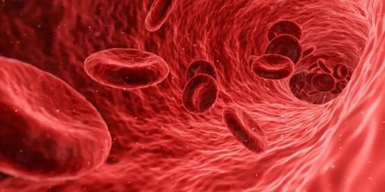
Red Blood Cells in Human Blood Vessels |
First tracking of microrobots
“Precise visualization and tracking of microrobots is necessary before they can actually serve humans,” says Paul Wrede, a doctoral researcher at the Max Planck-Zurich Center for Learning Systems Research, lead author of the paper.
“Without the aid of imaging technology, microrobots are approximately blind. Therefore, real-time, high-resolution imaging is essential for the detection and manipulation of microrobots the size of cells in living organisms.” Member of the Max Planck-Zurich Center for Learning Systems Research, Confederation of Zurich Daniel Razansky, professor of biomedical imaging at the Institute of Technology and the University of Zurich, added. In addition, imaging technology is also a prerequisite for monitoring robots performing treatments and verifying their task completion. “Therefore, if microrobots are to be used in the clinic, imaging systems need to be developed to track them in real time.”
In a recent study published in Science Advances , the team showed that they successfully used non-invasive imaging to clearly detect and track microscopic 5-micron-sized microscopic vessels in the brain blood vessels of mice in real time. robot. Together with Metin Sitti, a world-leading expert in microrobots, director of the Max Planck Institute for Intelligent Systems (MPI-IS), professor of physical intelligence, and other researchers, the team has achieved significant results in the effective combination of microrobots and imaging techniques breakthrough.

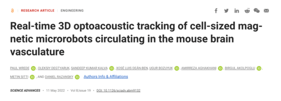
webpage Screenshot
The researchers used microrobots ranging in size from 5 microns to 20 microns, the smallest of which are about the size of red blood cells, 7 to 8 microns in diameter. Microrobots of this size can penetrate even the tiniest microcapillaries in a mouse brain when injected intravenously.
At the same time, the researchers have also developed a dedicated photoacoustic tomography technique to continuously monitor robots entering hard-to-reach areas deep in the body and brain in real-time and at high resolution, something that cannot be done with light microscopy or any other imaging technique. arrived. This method, known as photoacoustic imaging, produces high-resolution volumetric reconstructed images because light can be emitted and absorbed by various tissues, and the tiny ultrasound waves generated after absorption can be used for detection and analysis.
“Double-faced” Janus Robot
In order for the microrobots to be clearly visible in the images, the researchers needed to find a suitable contrasting material. They used microrobots made of spherical silicon microparticles with a Janus-like coating. Janus is the door god and protector in ancient Roman mythology, also known as the double-faced god. Scientists have taken inspiration from this to make the two sides of the microrobot spheres coated differently, half nickel and half gold. This type of robot has a sturdy structure and is ideal for performing complex medical tasks.

The spherical microrobots consist of silica-based particles, half coated with nickel (Ni) and the other half with gold (Au), and loaded with green-stained nanobubbles (liposomes). In this way, they can be detected individually using a new photoacoustic imaging technique | ETH Zurich/MPI-IS
The “double-sided” structure of microrobots
“Gold is a very good contrast material for photoacoustic imaging,” explains Razansky. “Without the gold layer, the signals from the microrobots are too weak to be detected.” In addition to gold, the researchers Microbubbles called nanoliposomes, which contain a fluorescent green dye, are also tested and can also be used as contrast agents. “An additional advantage of nanoliposomes is that they can be loaded with drugs, which could help in future drug-targeted therapy,” Wrede added. The potential use of liposomes will be further investigated in future work.

Microrobots in mouse blood vessels observed one by one with photoacoustic imaging | ETH Zurich / Max Planck Institute for Intelligent Systems
In addition, the gold coating also minimizes the cytotoxicity of the nickel coating—after all, if future microrobots are to operate in living animals or humans, they must be biocompatible and non-toxic. In the current study, the researchers used nickel as the magnetic actuation medium and used a single permanent magnet to steer the robot. In a follow-up study, they hope to test photoacoustic imaging by using rotating magnetic fields for more sophisticated manipulations.
“Follow-up research will focus on probing how to precisely manipulate microrobots in a state of rapid blood flow,” said Metin Sitti. “In the current study, we mainly focus on the visualization of microrobots. This project has achieved a major breakthrough, thanks to the The excellent collaborative environment at CLS allows the combined expertise of the robotics research group at MPI-IS in Stuttgart and the imaging research group at ETH Zurich.”
references
[1] https://ift.tt/k4Utybs
[2] https://ift.tt/pJY3BNz
Compilation: Forty Seven
Editor: Jin Xiaoming
Typesetting: Yin Ningliu
Source of the title map: Reference [1]
research team
Corresponding author Daniel Razansk: Professor, Department of Biomedical Imaging, Department of Information Technology and Electrical Engineering, University of Zurich Medical School and ETH Zurich
First author Paul Wrede: PhD researcher, Physical Intelligence, Max Planck-Zurich Research Center for Learning Systems
Research group website
https://ift.tt/fXtI6Ui
https://ift.tt/IvKq1k8
Paper information
Published the journal Science Advances
Posted on May 11, 2022
Title of the paperReal-time 3D optoacoustic tracking of cell-sized magnetic microrobots circulating in the mouse brain vasculature
(DOI: https://ift.tt/5SCpbxB)
The Future Light Cone Accelerator is an early-stage technology entrepreneurship accelerator initiated by Nutshell Technology. It provides scientists with solutions at different stages ranging from company registration, intellectual property rights, to financing needs, and team formation. Accelerate the transformation of scientific and technological achievements from the laboratory to the market, and accelerate the iteration of some scientists to become CEOs.
The Nutshell team has 12 years of experience in serving scientists. We always make suggestions from the perspective of scientists and be good friends of science and technology creators. If you are planning to start a technology business, whether you are looking for money, people, resources, or orders, you are welcome to chat with the Future Light Cone team. You can send bp or other project information to [email protected] , and leave your contact information, or add the Wechat of Guoke Hard Technology Enterprise to communicate by private message.

✦
✦
Click to read the original text to view the original paper
This article is reproduced from: http://www.guokr.com/article/461645/
This site is for inclusion only, and the copyright belongs to the original author.